Cookie Policy: This site uses cookies to improve your experience. You can find out more about our use of cookies in our Privacy Policy. By continuing to browse this site you agree to our use of cookies.
AdipoGen Life Sciences
anti-SARS-CoV-2 Spike Protein S1 (NTD), mAb (rec.) (AB72-1-H10)
| Product Details | |
|---|---|
| Synonyms | SARS-CoV-2 Spike S1 N-Terminal Domain Protein |
| Product Type | Recombinant Antibody |
| Properties | |
| Clone | AB72-1-H10 |
| Isotype | Mouse IgG2a |
| Source/Host | Produced without the use of animals. Purified from HEK 293 cell culture supernatant. |
| Immunogen/Antigen | Recombinant SARS-CoV-2 S1/S2 Protein (aa 14-1208 (proline substitutions at residues 986 and 987; “GSAS” substitution at the furin cleavage site (residues 682–685)) containing a C-terminal His-tag. |
| Application |
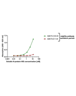
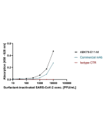
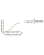
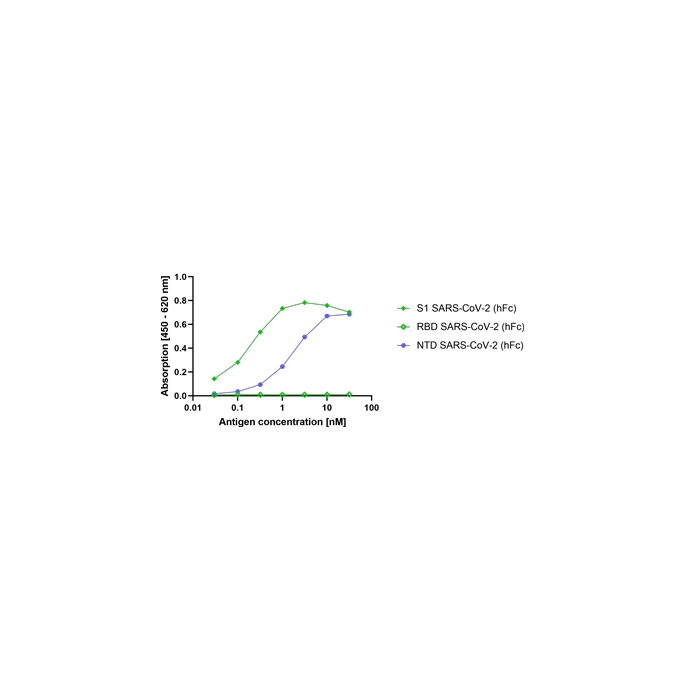
ELISA. Works as a capture antibody when coated on the immunoassay plate. Can be used together with the detection antibody AG-27B-6310. Does not work in a setup where the spike protein is directly coated on the immunoassay plate. |
| Crossreactivity | Virus |
| Specificity |
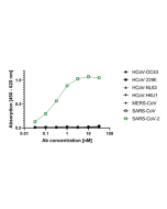
Recognizes the SARS-CoV-2 S1 (N-terminal domain). Does not cross-react with HCoV-OC43, HCoV-229E, HCoV-NL63, HCoV-HKU1, MERS-CoV or SARS-CoV. |
| Purity | ≥95% (SDS-PAGE) |
| Purity Detail | Protein A purified from animal component-free supernatant. |
| Concentration | 1 mg/ml |
| Formulation | Liquid. In PBS. |
| Isotype Negative Control | |
| Other Product Data |
This is an antibody developed by antibody phage display technology using a human antibody gene library from convalescent COVID-19 patients. For this antibody both the heavy and light chains are cloned and expressed, generating full-length antibodies. It has a Y-shaped structure of a common full-length IgG, consisting of 4 polypeptide, 2 heavy-chains and 2 light-chains. |
| Accession Number | P0DTC2 (aa 1-319) |
| Declaration | Manufactured by Abcalis |
| Shipping and Handling | |
| Shipping | BLUE ICE |
| Short Term Storage | +4°C |
| Long Term Storage | -20°C |
| Handling Advice |
After opening, prepare aliquots and store at -20°C. Avoid freeze/thaw cycles. Please handle under sterile conditions to avoid contamination. |
| Use/Stability |
Stable for at least 1 year after receipt when stored at -20°C. Stable for at least 3 months after receipt when stored at +4°C. |
| Documents | |
| MSDS |
 Download PDF Download PDF |
| Product Specification Sheet | |
| Datasheet |
 Download PDF Download PDF |
Coronaviruses (CoVs) are enveloped non-segmented positive-sense single-stranded RNA viruses and can infect respiratory, gastrointestinal, hepatic and central nervous system of human and many other wild animals. Recently, a new severe acute respiratory syndrome β-coronavirus called SARS-CoV-2 (or 2019-nCoV) has emerged, which causes an epidemic of acute respiratory syndrome (called coronavirus human disease 2019 or COVID-19). SARS-CoV-2 shares 79.5% sequence identity with SARS-CoV and is 96.2% identical at the genome level to the bat coronavirus BatCoV RaTG133, suggesting it had originated in bats. SARS-CoV-2 contains 4 structural proteins, including Envelope (E), Membrane (M), Nucleocapsid (N) and Spike (S), which is a transmembrane protein, composed of two subunits S1 and S2. The S protein plays a key role in viral infection and pathogenesis. The S1 subunit contains the N-terminal domain (NTD) and a receptor binding domain (RBD), which binds to the cell surface receptor Angiotensin-Converting Enzyme 2 (ACE2) present at the surface of epithelial cells, causing mainly infection of human respiratory cells, whereas S2 harbors heptad repeat 1 (HR1) and HR2. The RBD domain first binds its receptor to form an RBD/ACE2 complex. This triggers conformational changes in the S protein, leading to membrane fusion mediated via HR1 and HR2 and consequently in viral entry into target cells. Antibodies targeting various regions of S protein have different mechanisms in inhibiting SARS-CoV-2 infection. For example, NTD-targeting antibodies bind the NTD to form an NTD/mAb complex, thereby preventing conformational changes in the S protein and blocking membrane fusion and viral entry. RBD-targeting antibodies form RBD/mAb or RBD/Nb complexes that inhibit binding of the RBD to ACE2, thereby preventing entry of SARS-CoV-2 into target cells.


